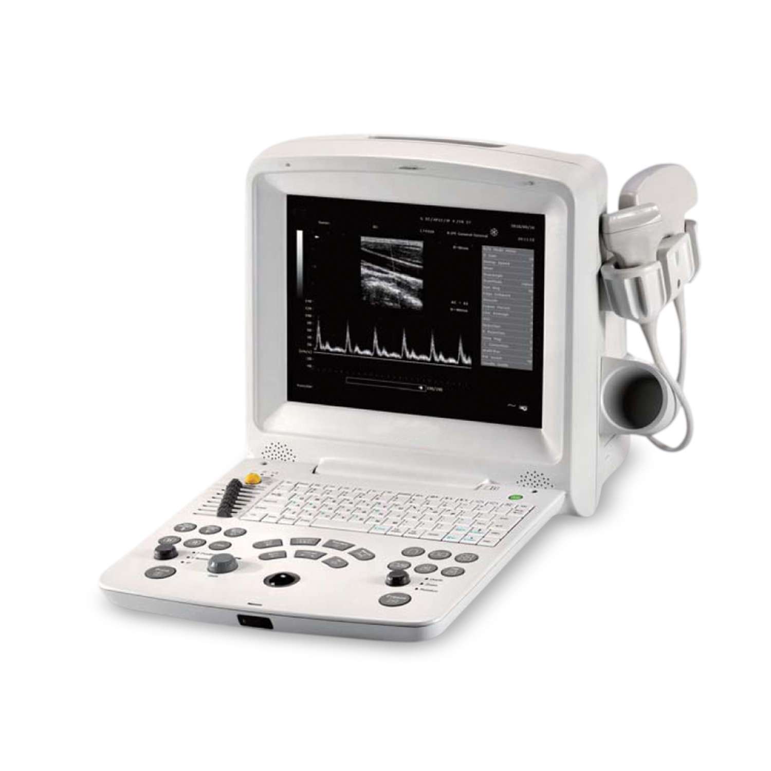
Powerful Technology
Complete digital beam forming technologies achieve high quality imaging
THI and TSI technology present sharp and clear imaging
PW Doppler brings more clinical diagnostic values on vascular disease
5 frequency broadband transducer selection for wide clinical applications
Compact and Portable
Compact and lightweight design for superior mobility
12.1″ folding high resolution TFT-LCD screen generates image clarity
Built-in battery ready for scanning two hours at point of care
User-Friendly Operation
One-touch image quality optimization by smart IP key
Backlit palm controller
User-defined keys contribute smooth operation
Quick-save keys for improved operation
Feasible Elements for Enhanced Operation
Intelligent 8-segment TGC for precise adjustment
Multi-format data transferring via USB and DIACOM
Multiple color enhancement options for personalized needs
Options
Linear array transducer L743UA (6.0/7.0/8.0/9.0/10.0MHz)
Linear array transducer L763UA (6.0/7.0/8.0/9.0/10.0MHz)
Convex array transducer L343UA (2.0/3.0/4.0/5.0/6.0 MHz)
Micro-convex array transducer C321UA(2.0/3.0/4.0/5.0/6.0 MHz)
Micro-convex array transducer C613UA(4.5/5.5/6.5/7.5/8.5 MHz)
Endorectal transducer E743UA (6.0/7.0/8.0/9.0/10.0MHz)
Endovaginal transducer E613UA (4.5/5.5/6.5/7.5/8.5MHz)
Needle-guided brackets for transducers
Also available: Video printer, laser printer,DICOM3.0, Footswitch
Mobile trolley, carrying bag, Lithium Battery
DRE FS-60 Advance Digital Ultrasound Machine
Affordable digital ultrasonic diagnostic imaging system features enhanced support of PW imaging
Powered by innovative technology, the DRE FS-60 provides optimal ultrasonic images. It has a maximum of 128 frames of built-in storage and a standard configuration of two transducer-connectors, giving you greater flexibility. The DRE FS-60 also has features typically exclusive to higher-end systems
General
Scanning angle From 30 to 155 degree(depending on transducers)
Scanning depth (mm) From 20 to 250(depending on transducers)
Imaging mode B, B+B, 4B, B+M, M and PW
Gray scales 256
Display 12.1″ TFT-LCD
Transducer frequency 2.0 ~ 10MHz
Transducer connector 2 standard
Beam-forming:
Digital beam-forming
Dynamic receiving focusing
Real-time dynamic aperture
Dynamic frequency scanning
Dynamic apodization
Tissue harmonic imaging
Tissue specific imaging
Imaging Processing
Pre-processing:
Dynamic range
Edge enhancement
Frame correlation
Line correlation
Smooth
AGC
8-segment TGC adjustment
IP (image process)
Post-processing:
Gray map
Gamma correction
Rejection
Colorization
Left-right reverse
Up-down reverse
Functions
Cine loop: 256 frames bidirectional cine-loop
Zoom: X1.0, X1.2, X1.4, X1.6,X2.0, X2.4, X3.0, X4.0 in real time
Storage media: Built-in flash,external USB-memory stick
Built-in image achive: 504 MB built-in image storage
Body mark: >80 types
Transducer: Auto detection
16-segment acoustic power output adjustment
Measurement & Calculation
B-mode: Distance, circumference, area,volume, ratio, stenosis%, and angle
M-mode: Distance, time, slope and heart rate
D-mode: Time, heart rate, velocity, acceleration, trace and RI
Software packages: abdomen, gynecology,obstetrics, urology,small parts, cardiology,
Software packages: orthopedics, cardiology, periperal vessels, and urology
Display
Date, time, probe name, probe frequency, frame rate, patient name, patient ID,
Hospital name, measurement values, body marks, annotation, probe position, full-image-regionedit
Standard Configurations
12.1″ TFT-LCD monitor
Two transducer connectors
256 frames cine loop memory
504 MB built-in image storage
Two USB ports
Measurement and calculation software packages
Convex array transducer C363-1 (2.5/3.5/5.0MHz)
